
The 2026 Nobel Prize in Medicine
Mary Cundiff, Ph.D.
November 2026
(11 Minutes)
If you would like to learn more about Alfred Nobel and the process behind these prizes, check out this article I wrote about last year’s winners. This year, some amazing discoveries received their well-deserved recognition.
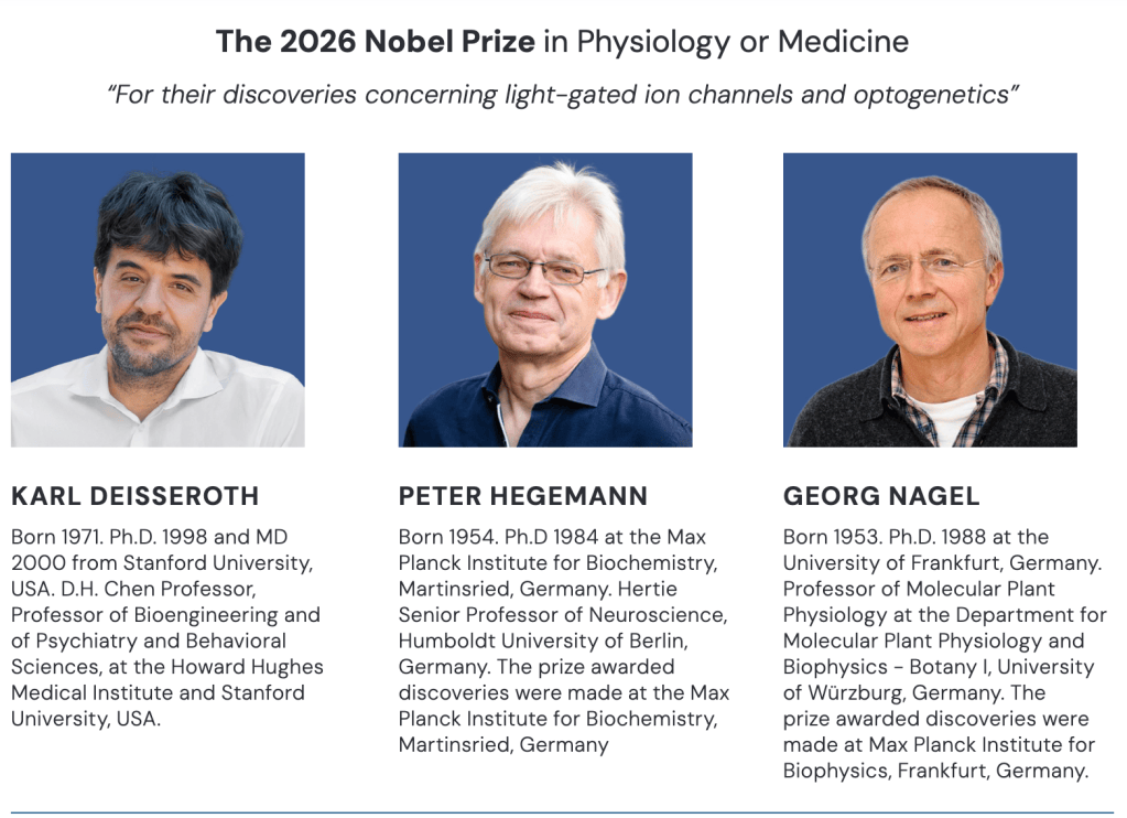
This article will focus on the 2026 Nobel Prize in Physiology or Medicine, awarded to Peter Hegemann, Georg Nagel, and Karl Deisseroth for their discoveries concerning light-gated ion channels and optogenetics.
As someone who spent my entire PhD performing optogenetic experiments in mouse brains, I literally jumped out of bed when I saw that optogenetics had finally won this prestigious award.
And the story behind its discovery is just as fascinating as the science itself.
What Is Optogenetics?
What is optogenetics, and how did it turn into something resembling “mind control”?
The world of neuroscience has long been interested in understanding the mysteries of human thought. How do we form memories? What happens in our brains when we daydream? How can a collection of cells produce something as complicated as creativity, emotion, or consciousness?
More importantly, how can we even begin to measure these things?
We know that neurons communicate through electrical signals called action potentials, which can occur in just milliseconds. Researchers have developed remarkable methods for recording these signals, but understanding whether a specific population of neurons actually causes a particular behavior is a much more difficult problem.
For decades, scientists relied on electrical stimulation or pharmacological interventions to manipulate neuronal activity. However, electrical stimulation can activate many nearby neurons, while drugs often act too slowly or affect too many cells to provide precise control. Ideally, researchers needed something that could activate specific neurons at the same speed they naturally communicate. And what could possibly be faster and more precisely controlled than light?
Interestingly, the answer came not from studying the human brain, but from studying algae.
The Curious Case of Light-Seeking Algae
In the 1990s, Peter Hegemann, working at the Max Planck Institute in Germany, was fascinated by a phenomenon known as phototaxis, the ability of an organism to move in response to light.
Specifically, he was interested in a single-celled green alga called Chlamydomonas. These tiny organisms have an incredible ability to detect and swim toward light. This can be demonstrated by placing the algae in a dish and illuminating one side. Within a short period, the algae begin accumulating toward the light source (Figure 1).
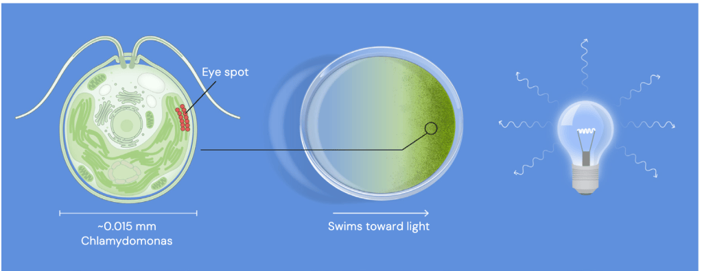
Why would algae need to do this? Like plants, algae perform photosynthesis, converting light energy into chemical energy. Being able to locate a suitable light source is therefore incredibly important for their survival.
Chlamydomonas has a specialized structure called an eyespot, which helps it determine the direction of incoming light. Although much simpler than the human eye, this structure contains light-sensitive proteins that allow the organism to respond to its environment. Interestingly, the human eye uses a related type of light-sensitive protein called rhodopsin.
In our retinas, light activates proteins that initiate a molecular signaling cascade. This eventually changes the activity of ion channels, which regulate the movement of electrically charged particles across cell membranes. But Hegemann noticed something remarkable about the algae: their electrical response to light was extraordinarily fast.
How could such a simple organism respond to light so rapidly?
He suspected that the answer involved a different molecular mechanism. Rather than relying on an extended series of molecular interactions, perhaps the algae possessed a protein that could directly convert light into an electrical signal. If true, this would mean that the light-sensitive protein and the ion channel were essentially combined into a single molecular machine. The challenge was identifying that protein.
Discovering the Molecular Switch
After years of investigating the algae’s light response, Hegemann’s team identified two promising genes in 2001 that resembled genes encoding other light-sensitive proteins. But identifying a gene is one thing. Understanding what its protein actually does is another.
To determine whether these genes encoded light-sensitive ion channels, Hegemann collaborated with biophysicist Georg Nagel, who specialized in studying the electrical properties of proteins. And this is where frogs entered the story. Nagel used immature egg cells, called oocytes, from the African clawed frog (Xenopus laevis) as an experimental system.
Why frog eggs?
These cells are relatively large and excellent for studying individual proteins. Researchers can introduce genetic material into the eggs, allowing them to produce a specific protein and incorporate it into their cell membranes. Electrodes can then be used to measure electrical currents across those membranes. Essentially, frog eggs provide a convenient experimental system for investigating how an unfamiliar protein works.
Nagel introduced the algal genes into frog eggs and exposed the cells to light. Remarkably, the eggs produced electrical currents in response to illumination. The researchers had identified proteins that could directly open an ion channel when exposed to light!
In 2002, they published their discovery of channelrhodopsin-1 (ChR1). The following year, they characterized a second protein called channelrhodopsin-2 (ChR2).
ChR2 was particularly exciting.
When exposed to blue light, the protein changed its structure, opening a channel that allowed positively charged ions to cross the cell membrane. This movement of charged particles altered the electrical properties of the cell, a process called depolarization. This is precisely the type of electrical change that can trigger an action potential in a neuron.
The researchers had discovered a protein that could potentially turn an ordinary cell into a light-sensitive cell. And perhaps most importantly, the protein worked outside of algae. Now someone just needed to introduce it into neurons.
From Algae to the Brain
Meanwhile, across the Atlantic, a neuroscientist and psychiatrist named Karl Deisseroth was interested in understanding how specific neural circuits contribute to behavior and psychiatric disorders. One of the greatest limitations in neuroscience was the inability to manipulate individual populations of neurons with sufficient precision.
The brain contains billions of neurons, all communicating through complicated networks. Even within a small brain region, different populations of neurons can have completely different functions. Researchers needed a way to distinguish which neurons were responsible for specific behaviors.
When Deisseroth and his collaborators learned about Nagel and Hegemann’s discovery, they recognized its extraordinary potential. What if they could take the gene encoding ChR2 from algae and introduce it into neurons? Would those neurons then respond to blue light?
In 2004, Deisseroth’s team, including Edward Boyden and Feng Zhang, began investigating this possibility. They obtained the ChR2 gene and introduced it into cultured mammalian neurons. Amazingly, the neurons successfully produced the protein and incorporated it into their membranes. Even more remarkably, when the researchers exposed the cells to brief pulses of blue light, the neurons fired action potentials.
Not only could they activate the neurons, but they could also control the timing of their electrical activity with millisecond precision. In 2005, the team published their groundbreaking findings, demonstrating that neuronal activity could be directly controlled using light.
Optogenetics had arrived.
But there was still one major challenge. Activating neurons in a dish was one thing. How could researchers deliver light to neurons buried deep inside a living brain? The answer was a combination of genetic engineering and fiber optics. By introducing ChR2 into specific neuronal populations and implanting a thin optical fiber into the brain, researchers could deliver light directly to their cells of interest.
In 2007, Deisseroth’s team demonstrated this technique in living mice. By introducing ChR2 into neurons of the motor cortex and delivering blue light through an optical fiber, they could activate the neurons and produce whisker movements.
Think about that for a moment.
A protein discovered in algae could now be used to control movement in a mammal.
And the story didn’t stop there.
Researchers also adapted other light-sensitive proteins, including halorhodopsin, which could inhibit neuronal activity when exposed to yellow light. Now scientists could not only activate neurons but also silence them. They effectively had molecular ON and OFF switches for the brain.
So, Is This Really Mind Control?
Well, sort of!
Optogenetics does not allow scientists to read someone’s thoughts or remotely control a person’s mind. However, it does allow researchers to manipulate the activity of specific neurons and examine the consequences. And that is an incredibly powerful distinction.
The term “optogenetics” combines two essential components: optics, the use of light, and genetics, the ability to selectively express light-sensitive proteins in particular cells. The genetic component is especially important. Rather than activating every neuron within a brain region, researchers can use genetic techniques to target specific cell populations.
For example, imagine that two populations of neurons occupy the same brain region, but one promotes movement while the other suppresses it. If researchers stimulate the entire region electrically, they might activate both populations simultaneously, making it difficult to understand which neurons are responsible for the resulting behavior. With optogenetics, researchers can selectively introduce ChR2 into one population and activate those neurons independently. If stimulation produces a change in movement, they have evidence that this neuronal population can causally influence that behavior.
This is a fundamental shift in how neuroscientists study the brain. Rather than simply observing which neurons become active during a behavior, researchers can begin testing whether those neurons are actually responsible for producing it. Over the past two decades, optogenetics has helped scientists investigate the neural circuits underlying movement, sleep, anxiety, reward, memory, and countless other behaviors. It has also transformed our understanding of neurological and psychiatric conditions, including Parkinson’s disease, epilepsy, and depression.
From Basic Science to Human Health
Although optogenetics has primarily been used as a research tool, its potential applications extend far beyond understanding neural circuits. One particularly exciting area of research involves restoring vision. In certain forms of retinal degeneration, the cells responsible for detecting light become damaged or die. However, other neurons within the retina may remain functional.
Researchers are exploring whether introducing light-sensitive proteins into surviving retinal cells could restore some degree of visual responsiveness. Early clinical studies have provided encouraging evidence that optogenetic approaches may help restore limited visual function in people with severe vision loss. These approaches are still experimental, and directly applying optogenetics to the human brain presents considerable technical and ethical challenges.
Nevertheless, the technology continues to provide invaluable insights into the biological mechanisms underlying disease. And understanding those mechanisms is often the first step toward developing better treatments.
The Beauty of Scientific Curiosity
Perhaps my favorite part of this year’s Nobel Prize is how perfectly it illustrates the unpredictable nature of scientific discovery.
Peter Hegemann was not trying to develop a technique for controlling neurons in the mammalian brain. He was curious about how algae respond to light.
Georg Nagel was interested in understanding the electrical properties of an unfamiliar protein.
Karl Deisseroth wanted to develop better tools for investigating the complicated relationship between neuronal activity and behavior.
Three scientists working across different areas of biology helped connect a series of discoveries that ultimately revolutionized neuroscience. Of course, they were not working alone. Countless other scientists, including Suneel Kateriya, Ernst Bamberg, Edward Boyden, Feng Zhang, and many others, made important contributions to the development of optogenetics.
But that is one of the beautiful things about science. Discoveries rarely happen in isolation. They build upon decades of experimentation, collaboration, curiosity, and sometimes a little luck.
A seemingly simple question about why algae swim toward light eventually led to one of the most powerful tools ever developed for studying the brain.
And that is exactly why this discovery deserves a Nobel Prize. Not because scientists can now perform something resembling “mind control,” but because optogenetics fundamentally changed the questions we can ask about the brain and our ability to answer them.
Sometimes the discoveries that change the world begin with nothing more than someone being curious about how it works. And sometimes, the answer is algae.
Meet the Prize Winners!


Leave a Reply